상품상세정보
3D 아나토미 시리즈는
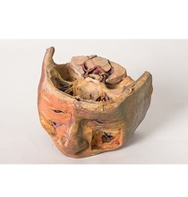
This 3D printed model captures a dissection in which the calvaria and cerebrum have been removed to expose the floors of the anterior and middle cranial fossae. The midbrain has been sectioned at the level of the tentorium cerebelli and on the cross sectional surface one can identify the superior colliculi, cerebral peduncles and the substantia nigra. Anterior to the mid-brain the vertebral artery can be clearly identified rising from the posterior cranial fossa and dividing into the posterior cerebral arteries. Anterior to this in the region of the sella turcica one can identify the internal carotid arteries emerging from the roof of the cavernous sinus medial to the anterior clinoid processes and beneath and lateral to the optic nerves and chiasm. The oculomotor nerves are visible penetrating the roof of the cavernous sinuses on the left and right posterior to the point where the internal carotid arteries emerge.
Anteriorly in the midline of the anterior cranial fossa lies the crista galli with the olfactory bulbs still present above the cribriform plates on either side. On the right the orbital plate of the frontal bone (the roof of the orbit) has been removed to expose the frontal nerve splitting into the supraorbital and supratrochlear nerves lying superior to the levator palpebrae superioris. The trochlear nerve is visible entering the superior aspect of the superior oblique muscle belly on the medial aspect of the orbit. Ethmoidal air cells have been exposed in the medial orbital wall by removal of the part of the lamina papyracea. On the left the levator palpebrae and superior rectus muscles have been divided along with the frontal nerve to expose the optic nerve, nasociliary nerve, ophthalmic artery and superior ophthalmic vein in the intraconal space.
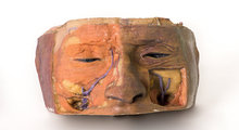
The face has been dissected to show facial muscles around the orbit on the right and the infraorbital nerve on the left. The infratrochlear nerve is also shown on the right and facial veins and arteries are also visible.
<번역>
이 3D 프린팅 모델은 두개골 앞쪽과 중간 부분의 바닥을 노출시키기 위해 대뇌와 대뇌가 제거된 절개를 포착합니다. 중간뇌는 천막소뇌 수준으로 분할되었고, 단면 표면에서 우량 결장, 뇌 경골, 그리고 상당한 흑질을 식별할 수 있다. 뇌 중간의 앞쪽에서 척추동맥은 두개골 후두에서 상승하여 후뇌동맥으로 분할되는 것을 명확히 확인할 수 있다. 셀라 터시카의 영역에서 이것의 앞부분은 동굴정맥의 지붕에서 전측 클리노이드 과정으로, 시신경과 염좌의 아래 및 측면으로 나타나는 내부 경동맥을 식별할 수 있다. 안구 운동 신경은 내부 경동맥이 나오는 지점으로부터 왼쪽과 오른쪽의 동굴 부비강 천장을 관통하는 것을 볼 수 있다.
두개골 앞부분의 중앙선 앞쪽에 크리스타갈리가 있고, 양쪽에 있는 크립리폼 판 위에 후각 구근이 여전히 존재한다. 오른쪽에는 전두골의 안와판(궤도의 지붕)이 제거되어 전두 신경이 안와와 상완골보다 위에 놓여 있는 초강력 신경으로 분열되는 것을 알 수 있다. 트로클리어 신경은 궤도의 안쪽 측면에 있는 상부 사선 근육의 윗부분으로 들어가는 것을 볼 수 있습니다. 유두엽의 일부를 제거함으로써 안쪽 안와벽에 에트모이드 공기 세포가 노출되었다. 왼쪽에는 전두신경과 함께 상완골근과 상완직근이 나눠져 시신경, 비강신경, 안동맥, 상안정맥 등이 교내공간에 노출됐다.
얼굴은 오른쪽의 궤도와 왼쪽의 안와 신경 주위에 얼굴 근육을 보여주기 위해 절개되었다. 오른쪽에도 인프라트로클리어 신경이 보이고 얼굴 정맥과 동맥도 보인다.


 보건교육지원센터를 시작페이지로!
  즐겨찾기
즐겨찾기