1523
Shoulder (left) Superficial muscles and axillary/brachial artery
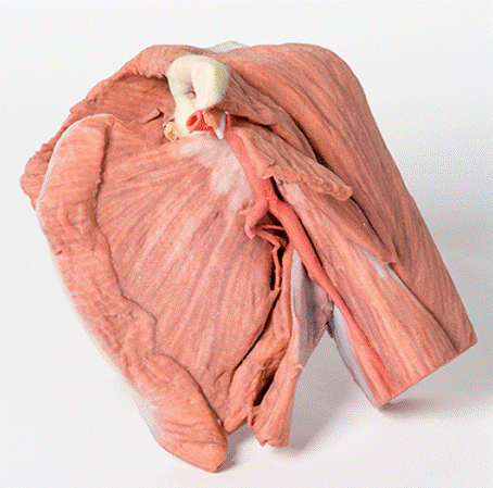
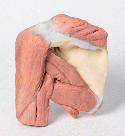
This printed 3D left shoulder specimen consists of the scapula, humerus (sectioned near midshaft) and clavicle (sectioned at midshaft) with the superficial muscles around the shoulder joint, the rotator cuff muscles and the axillary artery as it progresses distally to become the brachial artery.
The muscles attached to the clavicle have been preserved including the subclavius muscle attachment to the inferior border of the clavicle and the deltoid covering the lateral aspect of the proximal upper limb (overlying the origins of the long head of biceps brachii and the lateral head of triceps brachii). The clavicular head of the pectoralis major has been preserved. On the posterior aspect the superior fibers of trapezius can also be observed where they attach attached to the posterior border of the lateral third of the clavicle, and to the acromion process and the spine of the scapula. Other muscles attached to the scapula which have been preserved include the subscapularis and serratus anterior on the anterior or costal aspect. Inspection of the anterior aspect reveals that the pectoralis minor insertion onto the coracoid process of the scapula has been preserved. Posteriorly the teres major and teres minor muscles are clearly visible arising from the lateral border of the scapula. Supraspinatus is preserved but infraspinatus has partly been removed to show branches of the suprascapular artery passing from the supraspinous fossa around the base of the spine to enter the infraspinous fossa housing the infraspinatus muscle. A small part of the omohyoid attachment is also visible above the suprascapular ligament.
The axillary artery below the inferior border of the clavicle can be seen to give off the thoracoacromial branch anteriorly and just slightly more distally the suprascapular artery can be seen passing posteriorly. Coursing distally, it gives off posterior branches of the circumflex scapular and subscapular arteries. The anterior and posterior circumflex humeral arteries are hidden from view when viewed from in front, however the latter artery can be seen deep to the posterior fibres of deltoid as it emerges though quadrangular space. Below the inferior border of teres major the axillary artery becomes the brachial artery. The radial collateral artery is visible arising from the brachial artery. The axillary artery becomes the brachial artery beyond the lower margin of the teres major muscle.
The muscles of the proximal upper limb have all been preserved, and those of the superficial layer, i.e. long head of biceps brachii, and long and lateral heads of triceps brachii, can be observed to form a complete layer of musculature around the humerus. The cross section of the mid shaft of the humerus nicely displays the relations of the major neurovascular bundles and the muscles in the anterior and posterior compartments.
A small remnant of the suprascapular nerve passing under the suprascapular ligament is visible.
<번역>
이 3D 프린팅된 왼쪽 어깨 샘플은 어깨관절 주변의 표면 근육, 회전근육 및 겨드랑이 동맥이 상완동맥이 되도록 원위 방향으로 진행되면서 견갑골, 상완골(중축 부근 절개) 및 쇄골(중축 부근 절개)로 구성됩니다.
쇄골에 부착된 근육은 쇄골 하연에 부착되는 쇄골하근과 근위 상지의 측면 측면을 덮는 삼각근을 포함하여 보존되었다(이두근 상완골의 긴 머리와 삼두근 상완골의 측면 머리). 흉부전문의 쇄골두부는 보존되어 있다. 후방측면에서 사다리꼴의 상섬유는 쇄골의 외측 3분의 1의 후방 테두리, 견갑골의 견갑골 돌기와 척추에 부착되어 있는 곳에서도 관찰할 수 있다. 견갑골에 부착된 다른 근육들은 앞쪽 또는 늑골 측면의 앞쪽에 있는 견갑골하부와 톱니바퀴를 포함한다. 앞면을 검사한 결과 견갑골의 코라코이드 과정에 흉부 경미한 삽입이 보존되어 있는 것을 알 수 있다. 후방에는 견갑골의 외측 테두리에서 생기는 대근육과 소근육이 선명하게 보인다. 수프라스피나투스는 보존되어 있지만 척추의 기저부 주위의 수프라스피나투스와를 지나 수프라스피나투스를 수용하는 수프라스피나투스와로 들어가는 수프라스피나투스는 부분적으로 제거되었다. 오모하이드 부착물의 작은 부분도 견갑상 인대 위에서 볼 수 있다.
쇄골 하연 아래의 액와 동맥은 흉곽지방을 전방으로 발산하는 것을 볼 수 있으며, 조금 더 원위적으로 쇄골상동맥이 후방으로 지나가는 것을 볼 수 있다. 원위적으로, 그것은 견갑골과 견갑골하 동맥의 뒤쪽 가지를 발산한다. 전방 및 후방 상완골동맥은 전방에서 볼 때 시야에서 보이지 않지만 후자의 동맥은 사각 공간을 통해 나타나면서 삼각근의 후방 섬유까지 볼 수 있다. 대퇴골의 하연 아래에서 액와동맥은 상완동맥이 된다. 상완동맥에서 발생하는 요골측부동맥을 볼 수 있습니다. 겨드랑이 동맥은 주요 근육의 아래쪽 가장자리를 넘어 상완동맥이 된다.
근위 상지의 근육은 모두 보존되어 있으며, 상층의 근육, 즉 이두근의 긴 머리, 삼두근의 긴 머리, 그리고 횡두근의 긴 머리 등이 상완골 주위에 근육의 완전한 층을 형성하는 것을 관찰할 수 있다. 상완골 중앙축의 단면은 주요 신경혈관 다발과 전방 및 후방 구획의 근육의 관계를 잘 보여준다.
견갑상 인대 아래를 지나는 견갑상 신경의 작은 잔해가 보인다.


 보건교육지원센터를 시작페이지로!
  즐겨찾기
즐겨찾기


