1525
Shoulder - deep dissection of the left shoulder joint, musculature, and associated nerves and vessels
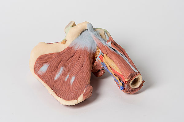
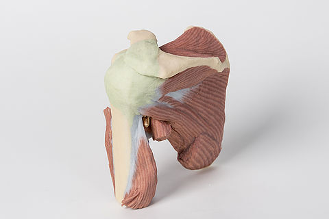
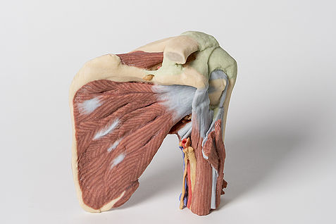
This 3D printed specimen presents a deep dissection of the left shoulder joint, musculature, and associated nerves and vessels of the scapula and proximal humerus (to near midshaft). Anteriorly, the deltoid muscle has been detached from its origin to expose the underlying deeper structures of the shoulder joint and rotator cuff musculature. The suprascapular nerve and artery are visible passing deep to, and superficial to, the superior transverse scapular ligament respectively. The multipennate subscapularis muscle is fully exposed with its tendinous insertion visible deep to the short head of the biceps brachii muscle. The insertion of the deltoid is preserved just overlying the long head of the biceps brachii, which ascends through the bicipital groove towards the glenohumeral joint capsule. Adjacent to the short head of the biceps brachii is the neurovascular bundle of the brachial artery, brachial vein, and terminal nerves of the brachial plexus (radial, ulnar, median, and the medial antebrachial cutaneous). The tendon of the latissimus dorsi, teres major, teres minor and long head of the triceps brachii muscles have been cut enhance the visibility of the medial aspect of the humerus, including the passage of the axillary nerve into the quadrangular space, the origin of the profunda brachii artery accompanying the radial nerve, and the insertion of the short head of the triceps brachii muscle. On the posterior aspect, the infraspinatus and supraspinatus muscles are fully exposed from their origins to insertions on the proximal humerus. The glenohumeral joint capsule is intact, with the extracapsular ligaments (e.g., acromioclavicular, coracoacromial, and coracoclavicular [both conoid and trapezoid portions]) preserved.
<번역>
3D 프린팅된 이 검체는 왼쪽 어깨 관절, 근육 구조 및 관련 신경과 견갑골 근위 상완골 혈관(중축 근처)의 깊은 절개를 보여줍니다. 앞에서, 어깨관절과 회전근개근의 밑바닥 더 깊은 구조를 드러내기 위해 삼각근은 원점에서 분리되었다. 견갑골상신경과 동맥은 각각 상부 횡격 견갑골 인대에 깊이 및 표면으로 전달되는 것을 볼 수 있다. 다년성 시상하근은 이두근의 짧은 머리 깊이까지 힘줄 삽입이 보이는 상태에서 완전히 노출된다. 삼각근의 삽입은 이두근을 통해 슬관절 캡슐을 향해 올라가는 이두근의 긴 머리 바로 위에 보존됩니다. 이두근 상완골의 짧은 머리 옆에는 상완동맥, 상완정맥 및 상완신경 말단신경의 신경혈관다발(반경, 척골, 중앙 및 내측 상완전피부)이 있다. 삼두근 상완근의 삼두근의 주요 근막, 경근, 경근 및 긴 두근의 힘줄이 절단되어 액와신경의 사각형 공간으로의 통과, 요골신경에 수반하는 상완골동맥의 원점 및 삽입을 포함한 상완골 내측면의 가시성이 향상되었습니다.상완 삼두근의 짧은 머리. 후측면에서는 근위 상완골에 삽입될 때까지 근위근과 근위근육이 완전히 노출되어 있다. 슬개골관절캡슐은 견갑외인대(예: 견갑골, 코라코크로미컬, 코라코클로미컬(원추상 및 사다리꼴 부분 모두))가 보존된 상태로 온전하다.


 보건교육지원센터를 시작페이지로!
  즐겨찾기
즐겨찾기