1530 Hand
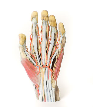
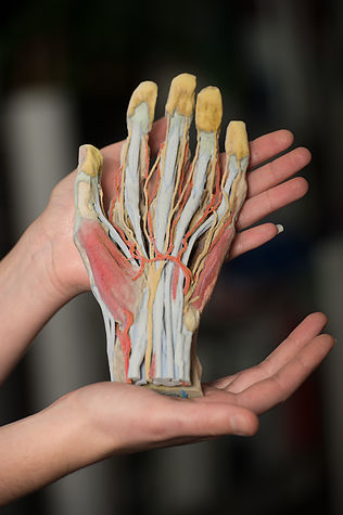
This 3D printed specimen demonstrates a superficial dissection of a left hand and wrist. Anteriorly, the transverse carpal and palmar carpal ligaments have been removed to expose the tendons and nerves traversing the carpal tunnel and Canal of Guyon. The palmar aponeurosis has been removed to demonstrate the course of the tendons through the palm, the superficial muscles of the thenar and hypothenar eminences (abductors and flexors), and the lumbrical muscles arising from the flexor digitorum tendon. In the digits, the fibrous sheaths have been removed to expose the flexor pollicis longus tendon and the spatial relationships between the flexor digitorum superficialis and profundus tendons as they insert into the intermediate and terminal phalanges. Also visible in the midpalm is the superficial palmar arch with contributions from superficial branches of the ulnar and radial arteries. The superficial palmar arch branches (common palmar) and terminal arteries (proper palmar digital) are visible to the terminal phalanges. Accompanying these vessels are the corresponding common and proper palmar digital nerves from the median and ulnar nerves. Also visible in the wrist are the tendons of the flexor carpi radialis and flexor carpi ulnaris tendons, and the radial and ulnar arteries.
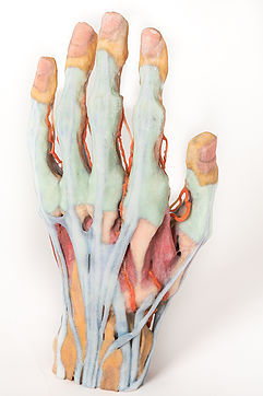
Posteriorly, the radial artery can be seen traversing the floor of the anatomical snuffbox and giving rise to both the deep branch (piercing the first dorsal interosseous muscle) and the dorsal carpal branch. The superficial fascia and extensor retinaculum has been removed to display the course and insertions of the extensor muscle tendons, as well as the tendons of the extensor pollicis longus, brevis, and abductor pollicis longus muscles. Both intertendinous connections and the extensor expansions (with insertions from the first dorsal interosseous and lumbrical) visible.
<번역>
이 3D 프린팅 시료는 왼손과 손목의 표면 절개를 보여줍니다. 앞에서는 손목 터널과 가이온 운하를 가로지르는 힘줄과 신경을 드러내기 위해 손목과 손목의 횡인대가 제거되었다. 손바닥을 통한 힘줄의 과정, 시너와 강하근의 표면 근육(유두와 굴곡), 그리고 굴곡 디지토럼 힘줄에서 발생하는 내강근의 과정을 보여주기 위해 팔뚝 무신경증이 제거되었다. 자릿수에서는 섬유질 시스가 제거되어 중간 및 말단 지골에 삽입될 때 굴곡성 폴리시스 롱우스 힘줄과 굴곡성 디지토리움 표피골 및 근막 힘줄 사이의 공간적 관계를 드러낸다. 척골과 요골 동맥의 표면 가지에서 기여하는 표면 팔마 아치도 중간 종려에서 볼 수 있다. 표면 손바닥 아치 가지(일반 손바닥)와 말단 동맥(적절한 손바닥 디지털)은 말단 지골에서 볼 수 있습니다. 이러한 혈관에는 중앙 신경과 척골 신경에 해당하는 일반적이고 적절한 장골 디지털 신경이 동반됩니다. 손목에 보이는 것은 요골 굴근과 척골 굴근의 힘줄과 요골 및 척골 동맥이다.
후방에, 요골 동맥은 해부학적 코담배 상자의 바닥을 가로질러 깊은 가지(첫 번째 골간 근육을 뚫는 것)와 등수근 가지 모두를 발생시키는 것을 볼 수 있습니다. 외피근막과 신장망막은 신장근 힘줄의 경로와 삽입을 나타내기 위해 제거되었으며, 신장근, 브레비스 및 외전근의 힘줄도 표시되었습니다. 간극 연결과 신장 확장(첫 번째 골간 및 내강 배면 삽입 포함)이 모두 보입니다.


 보건교육지원센터를 시작페이지로!
  즐겨찾기
즐겨찾기