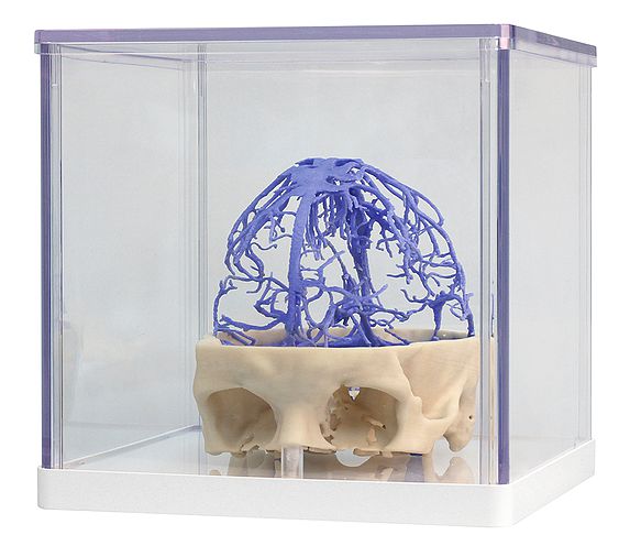
1645 Venous Circulation
This 3D print presents the same dataset that underlies our Circle of Willis and cranial arterial circulation 3D prints and is derived from careful segmentation of angiographic data. Here, the dural venous sinus network has been segmented based on structures visible from the circulation of contrast medium in the late phase of filling. As a result, while most of the sinuses are present, the lack of contrast in the anterior portions of the venous system means that some structures are not as clear in the model as may be expected – for example the cavernous sinus and inferior petrosal sinus.
The extensive network of dural veins and venous lacunae are visible, which drain towards the midline in the superior sagittal sinus. Deep to this network of sinus veins are the great cerebral vein which drains with the inferior sagittal sinus into the straight sinus which then converges with the superior sagittal at the confluence of sinuses. Several dural veins drain into the left and right transverse sinuses as they pass anteriorly towards the petrous portion of the temporal bone. The sigmoid sinuses can be seen in the posterior cranial fossa prior to exiting the skull at the jugular foramen and forming the internal jugular vein (visible on the inferior surface of the skull).
<번역>
이 3D 프린트는 Circle of Willis 및 Cranial 동맥 순환 3D 프린트의 기초가 되는 것과 동일한 데이터 세트를 제공하며 혈관 조영 데이터의 신중한 분할에서 파생됩니다. 여기에서 경막정맥동 네트워크는 충전 후기의 조영제 순환에서 볼 수 있는 구조에 기초하여 분할되었다. 그 결과, 대부분의 부비강이 존재하지만, 정맥계 앞부분의 대비가 부족하다는 것은 일부 구조(예: 동굴성 부비강과 하방성 부비강)가 예상만큼 모델에서 명확하지 않다는 것을 의미한다.
경막정맥과 정맥 라쿠나의 광범위한 네트워크가 보이는데, 상 시상정맥류는 시상정맥동의 중앙선을 향해 배수됩니다. 이 부비동정맥의 네트워크 깊은 곳에는 대뇌정맥이 있는데, 대뇌정맥은 하부 시상정맥과 함께 직정맥으로 흘러들어 부비동 합류점에서 상부 시상과 수렴한다. 몇 개의 경막정맥이 측두골의 페터러스 부분을 향해 전방으로 지나갈 때 좌우 횡부비강으로 빠져나갑니다. S자형 부비강은 경정골공에서 두개골로 나와 내부 경정맥을 형성하기 전에 두개골 후두와에서 볼 수 있습니다.


 보건교육지원센터를 시작페이지로!
  즐겨찾기
즐겨찾기