1665 Deep face/Infratemporal fossa
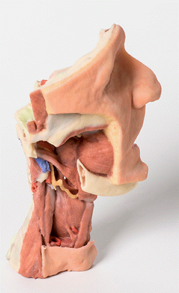
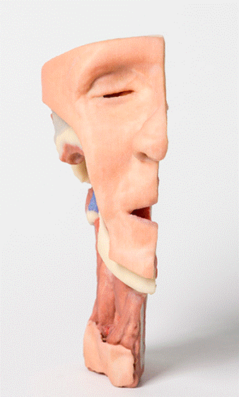
In this 3D printed specimen of a midsagittally-sectioned right face and neck, the ramus, coronoid process and head of the mandible have been removed to expose the deep part of the infratemporal fossa. The pterygoid muscles have also been removed to expose the lateral pteygoid plate and posterior surface of the maxilla. The buccinator has been retianed and can be seen originating from the external aspect of the maxilla, the pterygomandibular raphe and the external aspect of the (edentulous) mandible.The superior constrictor can also be seen arising from the posterior aspect of the pterygomandibular raphe. The internal laryngeal nerve has been preserved. Muscles in the neck that are identifiable include mylohyoid, the strap muscles and the inferior constrictor. The styloid muscles can be seen descending from the process to their insertions (not shown). The internal carotid artery can be seen deep to the styloid process which gives origin to stylohyoid, styloglossus and stylopharyngeus.
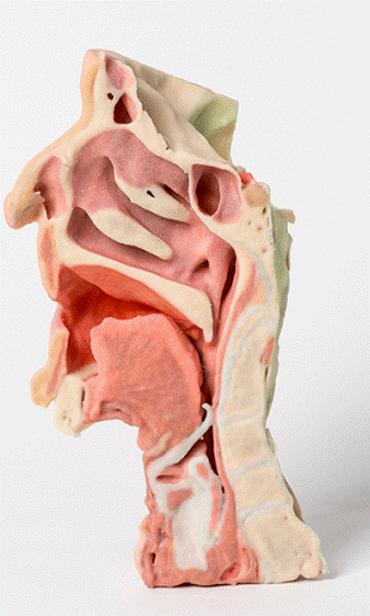
The sectioned surface preserves a series of midline head and neck structures, including: the lateral wall of the nasal cavity (superior, middle and inferior conchae and sphenoethmoidal recess, superior meatus, middle meatus and inferior meatus), the nasopharynx, the opening of the auditory tube, the hard palate, soft palate, the intrinsic tongue muscles, oropharynx, laryngopharynx, and hyoid bone. The parts of the laryngeal cartilages and the pharynx are clerly seen, as are the verterbral bodies of C2-C5, the anterior arch of C1(atlas), and the dens of C2 (axis).
<번역>
본 발명의 3D프린트 우측 얼굴과 목의 중간 절개 시료에서는 하악골의 라무스, 코로나이드 과정 및 두부를 제거하여 하악골 하악골의 깊은 부분을 노출시켰다. 익상근은 또한 상악골의 외측 익상판과 후면을 드러내기 위해 제거되었다. 볼시네이터는 수정되었으며 상악골, 익상악골 및 하악골의 외측면에서 유래한 것으로 볼 수 있다.상부 협착체는 익상악골 후측면에서도 볼 수 있다. 내부 후두신경이 보존되어 있습니다. 식별할 수 있는 목의 근육은 골수근, 스트랩근, 하부협착제 등이다. 스타일로이드 근육은 프로세스에서 삽입으로 내려오는 것을 볼 수 있습니다(표시되지 않음). 내경동맥은 스타일로이드, 스타일로그로스, 스타일인후두의 기원을 제공하는 스타일로이드 과정까지 볼 수 있습니다.
절개된 표면은 비강의 외벽(상부, 중부, 하부 원추 및 구상골 함몰부, 상부 미트, 중간 미트, 하부 미트), 비인두, 청각관 개구부, 경구개, 연구개, 내성 혀 뮤를 포함한 일련의 중앙선 머리와 목 구조를 보존합니다.사골, 구인두, 후두, 설골. C2-C5의 추골체, C1(아틀라스), C2(축)의 치밀과 같이 후두 연골과 인두의 일부가 잘 보인다.


 보건교육지원센터를 시작페이지로!
  즐겨찾기
즐겨찾기


