상품상세정보
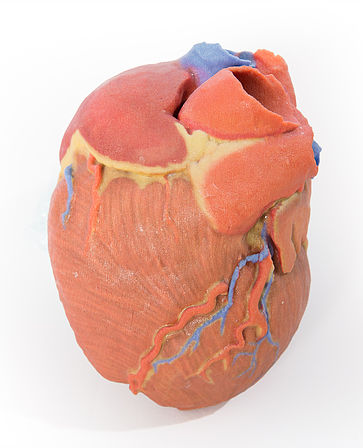
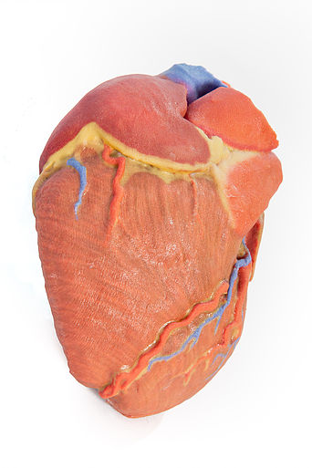
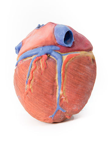
This 3D printed heart specimen preserves superficial cardiac anatomy and the bases of the great vessels. All four chambers (atria and ventricles) are preserved, with the pericardial reflections on the left atrium demarcating the position of the transverse and oblique pericardial sinuses. The right marginal branch of the right coronary artery is visible exiting from the fat-filled coronary sulcus, as well as the posterior interventricular (posteriro descending) artery within its sulcus. The anterior interventricular (left anterior descending) and diagonal branches from the left coronary artery are also visible anteriorly, as well as the terminal portion of the circumflex branch deep to the left auricle and great cardiac vein. On the posterior aspect, the coronary sinus receives all the cardiac veins (great, middle, small) and a prominent posterior vein of the left ventricle. The aortic and pulmonary semilunar valves are visible at the bases of the ascending aorta and pulmonary trunk, respectively.
<번역>
이 3D 프린팅 심장 검체는 표피적인 심장 해부학적 구조와 대혈관 밑면을 보존합니다. 좌심방의 심막 반사가 횡심막 및 사심막 부비강의 위치를 규정하는 4개의 챔버(심방 및 심실)가 모두 보존됩니다. 오른쪽 관상동맥의 오른쪽 주변 지점은 지방으로 가득 찬 관상동맥과 그 관상동맥의 후측심실(내림 후) 동맥에서 나오는 것을 볼 수 있다. 좌심실간(좌측 전방 하강) 및 좌관상동맥으로부터의 대각선 가지와 좌측 귓바퀴 깊숙이 있는 회음지 말단부 및 대심정맥도 전방에서 볼 수 있다. 후면에서, 관상동맥 부비동은 모든 심장 정맥(대, 중, 소)과 좌심실의 두드러진 후정맥을 받는다. 대동맥 및 폐반월판막은 각각 상승 대동맥과 폐간격의 기저부에서 볼 수 있습니다.


 보건교육지원센터를 시작페이지로!
  즐겨찾기
즐겨찾기